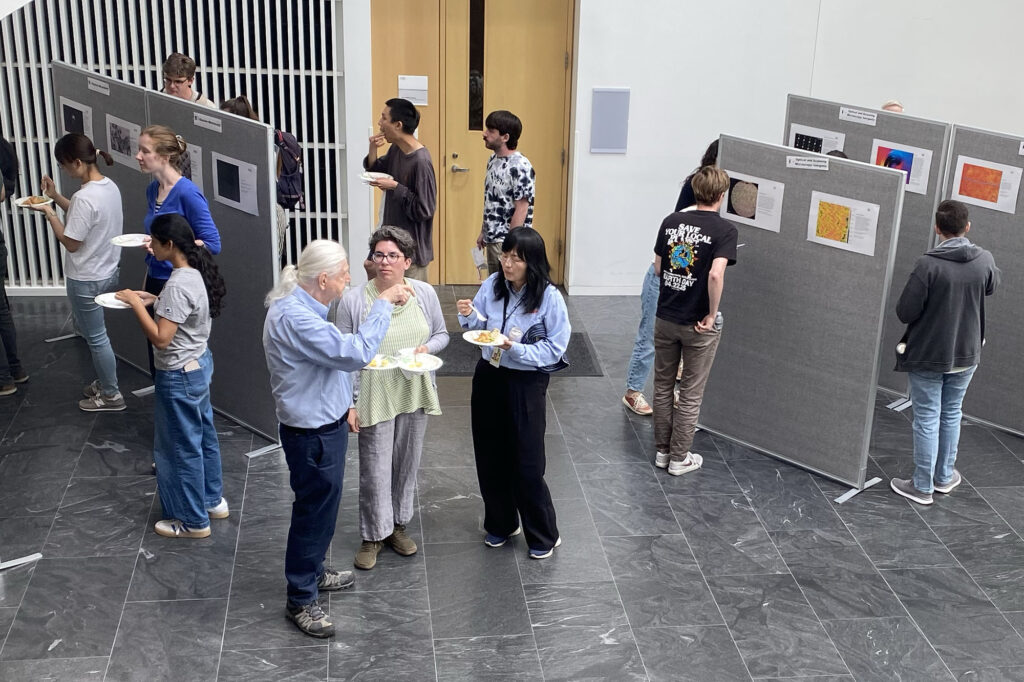
“Cobblestone That Gobbles Bone,” “The Sweetest Architecture,” and “Asymmetrical Eruptions on a Josephson Junction” sound more like movie titles than scientific research. They are, in fact, the titles of a few of the winning entries in the 2026 Cornell Imaging Contest, a campuswide competition that showcased the striking imagery researchers create using microscopes.
Supported by the Cornell Center for Materials Research (CCMR), the Biotechnology Resource Center (BRC), the Cornell NanoScale Science and Technology Facility (CNF), and the Student Council of the Microscopy Society of America, the contest received 57 submissions, and more than 150 people attended the Microscopy User Showcase on June 5.
For hundreds of Cornell researchers, microscopy is an essential tool, enabling discoveries in fields ranging from cellular biology and disease research to advanced materials, semiconductors, and quantum technologies.
Among the 57 submissions on display were images that revealed both the scientific power and artistic beauty of microscopy. The images are detailed, evocative, often visually striking, sometimes enhanced with artificial color for definition, or stained to show features of the material.
“In most cases, the images we capture are stored in our computers and are never seen by anyone else,” said Tathagata Banerjee, a doctoral student in applied phyics, and one of the organizers for this year’s competition. “This competition provided an opportunity for those images to be seen and appreciated by the community, and for us to share our research with others.”
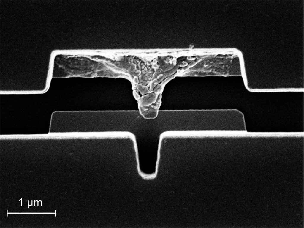
Banerjee’s image, “Asymmetrical Eruptions on a Josephson Junction” depicts a microscopic electronic device known as a Josephson junction, in which two superconducting electrodes are separated by a thin insulating layer. Applying high voltage to the junction caused one electrode to overheat and fail, but the rest of the device remained undamaged. The image won first place among the CNF submissions.
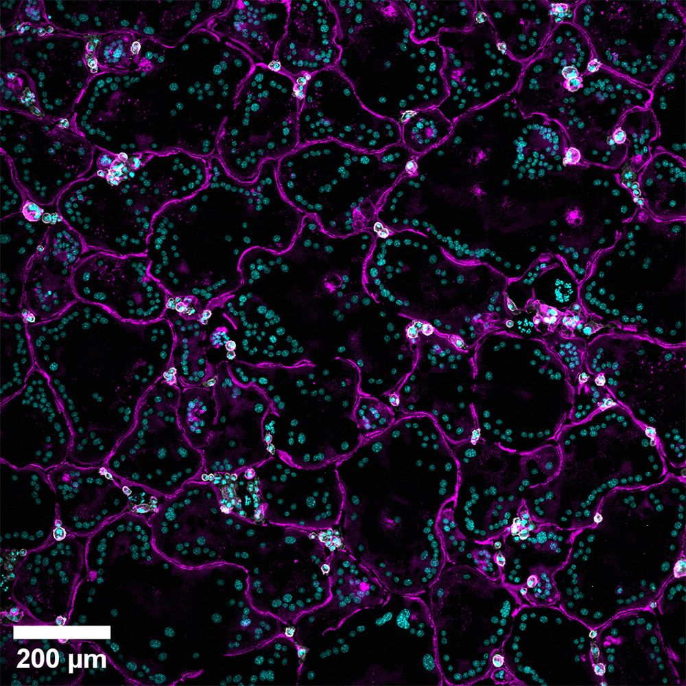
The first-place winner in the “Popular” category, “Cobblestone That Gobbles Bone” by doctoral student Kylie Persson, shows osteoclasts – cells that dissolve and remove damaged bone tissue – clustered together in a pattern suggesting a cobblestone road. In the image, the osteoclasts were fluorescently stained to highlight the various parts of the cell.
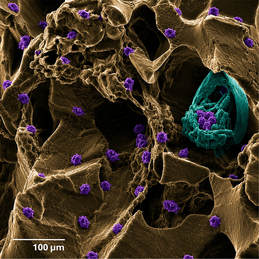
For some entries, the poetry of the caption matched the artistry of the image. Doctoral associate Shiseido Robinson created the image “The Sweetest Architecture” that won first place among CCMR submissions and second place in the “Popular” category. It depicts a porous structure created when sugar crystals were coated with a liquid silicone polymer (polydimethylsiloxane, or PDMS). After the sugar dissolved, it left behind a porous scaffolding where Jurkat cells – a laboratory-grown line of human immune cells – had attached and grown.
“Born from a sacrificial sugar template, this porous PDMS scaffold bears the fossilized memory of crystals that have long since dissolved,” Robinson wrote. “This scanning electron microscope image captures a paradox of creation through disappearance: the sugar vanishes, yet its imprint endures.” The work showed how microscopy can support immunotherapy research, by helping to design environments in which immune cells function more effectively against disease.
Reimagining an earlier vision
The contest builds on a tradition of microscopy image competitions at Cornell dating back to the early 2000s. In 2002, Cornell’s Department of Materials Science and Engineering hosted “Images in the Material World,” which was organized by Malcolm Thomas ’83, an electron microscopist and electron microscopy research facility manager at CCMR.
“I started the competition to highlight the work being done in our microscopy facilities,” said Thomas. “A second goal was to give students an opportunity to showcase their research, promote the science and their hard work, and help them work on the skills needed to communicate about why their work is important.”
The early competitions inspired Shake Karapetyan, a doctoral student in applied physics, and Naomi Pieczulewski, Ph.D. ’26 to revive the initiative in 2025. In 2026, with help from co-organizer Tathagata Banerjee and Jack Crowley, both Ph.D. candidates in applied physics, and Amanda Weiner, a Ph.D. candidate in biochemistry and molecular biology, the contest grew into a truly campuswide effort involving CCMR, BRC, and CNF as well as other departments and disciplines.
“One of our goals in starting the contest was to build a sustainable, student-led initiative,” Karapetyan said. “We wanted to celebrate the remarkable imaging being done across Cornell while also creating opportunities for researchers to connect with one another and learn about the imaging facilities available on campus.”
Building on a successful event in 2026, the organizers are working to expand the contest to attract participation from more disciplines across Cornell. They are also exploring ways to use the images to engage K-12 and undergraduate students while introducing prospective students to Cornell.
“These images are a powerful way to bring science and engineering research to younger students,” said Banerjee. “When we shared these images with high-school and undergraduate students, they were immediately interested in the science and excited to ask questions.”
The next Cornell Imaging Competition is planned for late spring 2027, with organizers encouraging submissions from across the university starting mid-April. The organizers also credited the staff across Cornell’s facilities for their support and encouragement, particularly Alexandra Pfeifer, Jon Shu, Vivek Iyer, and Rebecca M. Williams, among many others at CCMR, BRC, and CNF. Their contributions helped make the event become the campus-wide collaboration envisioned by the organizers.
Microscopy at Cornell consistently pushes the limits of innovation with development of advanced equipment. In April 2026, Professor David A. Muller, Ph.D. ’96, Samuel B. Eckert Professor of Engineering in the School of Applied and Engineering Physics, was elected to the National Academy of Engineering for his contributions to developing the highest resolution electron microscope in the world.
“Microscopy allows us to see structures and processes that cannot be viewed in other ways,” said Muller, who is also co-director CCMR and the Kavli Institute at Cornell for Nanoscale Science. “By continuously pushing the boundaries of what microscopy can do, Cornell is helping researchers explore everything from living cells to quantum materials, creating images that spark curiosity and inspire awe, and providing students access to some of the world’s most advanced imaging tools.”